Monday, 16 May 2016

Case #3


Clinical History :

19 years old male patient with history of SOB.


------------------------------------------------------------------------------------------------


Findings:


There is a faint left lower lobe retrocardiac opacity (red arrows) associated left plural effusion ( yellow arrow).
Left pigtail (blue arrow).


------------------------------------------------------------------------------------------------


Enhanced chest CT scan was performed


------------------------------------------------------------------------------------------------


FINDINGS


There is tabulated highly vascularized left para-spinal mass( red arrow) showing heterogeneous peripheral enhancement with necrosis and a focus of calcification centrally ( blue arrow). There is large feeding vessels ( green arrow) connecting to a very large aneurysmally dilated para-spinal vessel ( orange and white arrows). No intraspinal extension. 


--------------------------------------------------------------------------------------------------------------


Final diagnosis :

Neuroendocrine tumor.


Case #2


  History :

29 years old male patient with history of chest trauma 


-----------------------------------------------------------------------------------------------------------


Findings: 


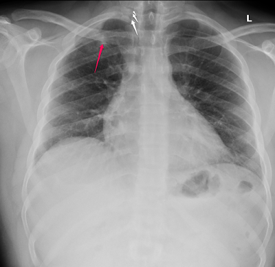
There is right 1st rib fracture with evidence of superior pneumomediastinum ( white arrows), also there is  small opacity seen in the right apex ( red arrow) given the patient's history this likely represent contusion. Further evaluation by CT scan is advised.


------------------------------------------------------------------------------------------------------------


------------------------------------------------------------------------------------------------------------


Findings :


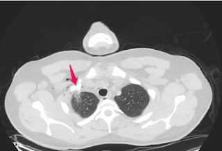
There is superior pneumomediastinum corresponding to the findings on the chest x-ray


Also there is subcutaneous emphysema.


There is a  displaced fracture of the first rib.


The lung apex shows an opacity in the right upper lobe adjacent to the fracture likely represent a contusion.


Saturday, 14 May 2016

Case #1

27 years old female patient with history of chronic dysphagia


This PA chest x-ray demonstrate an air-fluid level in the superior mediastinum likely arising from the esophagus, the distal esophagus is no dilated. The lung grossly appears clear 


DDx include :

1- Esophageal diverticulum.
2- Infected larynogcele.
3- Infected cyst.


 Enhanced CT scan has been performed 


This an enhanced CT scan at the level of the aortic arch demonstrate an out-pouching arising from the cervical part of the esophagus, no adjacent lymph nodes. Otherwise no gross abnormality could be identified.

The findings are in keeping with esophageal diverticulum(Zenker diverticulum).


Types of esophageal diverticulum:


1- Zenker’s Diverticulum :


Pharyngoesophageal diverticulum, located at the posterior superior aspect at the thyropharyngeal muscle and inferior boundary is cricopharyngeal muscle.


2- Midesophageal Diverticulum


3- Epiphrenic Diverticulum:

Located at the lateral distal part of the esophagus more common on the right side.