
Wednesday, 18 January 2017

Saturday, 5 November 2016
Case #1
History:
A 39-year-old female with history of chest pain.


Findings:
There is a large left lung bullae occupying more than half of the lung.
Diagnosis:
Vanishing lung syndrome
case #4
History:
A 18-month-old girl with chest wall mass, for evaluation.


Findings:
There is a left lateral chest wall subcutaneous lesion. The lesion is hyperintense on T1 and T2 and low signal in fat-sat sequence. No definite soft tissue enhancing component.
Diagnosis:
Lipoblastoma
case #3
History:
36 months old with right thigh swelling.


Findings:
The x-ray shows swelling lateral to the right femur with no evidence of bone disruption, infiltration or lytic changes.
The MRI shows large well-defined lobulated soft tissue mass noted arising from the lateral femoral compartmental muscles. It shows a well-defined capsule and central necrosis. The lesion is isointense to muscle in T1 weighted images and high in T2 weighted images with heterogeneous enhancement. The adjacent femoral vessels are compressed by the mass.
Diagnosis:
Rhabdomyosarcoma
Case #2
Known case of prune belly syndrome and the image is shown
just to see the appearance of the disease.

Case #1
History :
No history was given


Findings:
A well-defined expansile bony lesion arising from the left transverse process of T7 vertebral body is noted extending posteriorly to the chest wall displacing the left erector spinae muscle posteriorly.
Diagnosis:
osteochondroma
Friday, 21 October 2016
Case #2
History:
79 years old male patient with history of left lower abdominal pain.

Findings:
The left lower abdomen demonstrates fatty lesion surrounded by hyperdense rim and fat strandings.
Diagnosis:
Acute Epiploic Appendagitis
Case #1
History :
77 years old male patient with history of abdominal pain.


Findings:
In the arterial phagse, both adrenal glands appear hyper-enhanced. In the porto-venous phagse the IVC appears collapsed.
In addition, there is a large intraperitoneal collection with enhanced wall that encapsulate the small intestine.
In addition, there is a large intraperitoneal collection with enhanced wall that encapsulate the small intestine.
Diagnosis:
These findings represent hypo-perfusion complex with large intraperitoneal collection.
Recommendation:
Monday, 16 May 2016
Case #3
Clinical History :
19 years old male patient with history of SOB.

------------------------------------------------------------------------------------------------
Findings:

There is a faint left lower lobe retrocardiac opacity (red arrows) associated left plural effusion ( yellow arrow).
Left pigtail (blue arrow).
------------------------------------------------------------------------------------------------
Enhanced chest CT scan was performed


------------------------------------------------------------------------------------------------
FINDINGS


There is tabulated highly vascularized left para-spinal mass( red arrow) showing heterogeneous peripheral enhancement with necrosis and a focus of calcification centrally ( blue arrow). There is large feeding vessels ( green arrow) connecting to a very large aneurysmally dilated para-spinal vessel ( orange and white arrows). No intraspinal extension.
--------------------------------------------------------------------------------------------------------------
Final diagnosis :
Neuroendocrine tumor.
Case #2
History :
29 years old male patient with history of chest trauma

-----------------------------------------------------------------------------------------------------------
Findings:
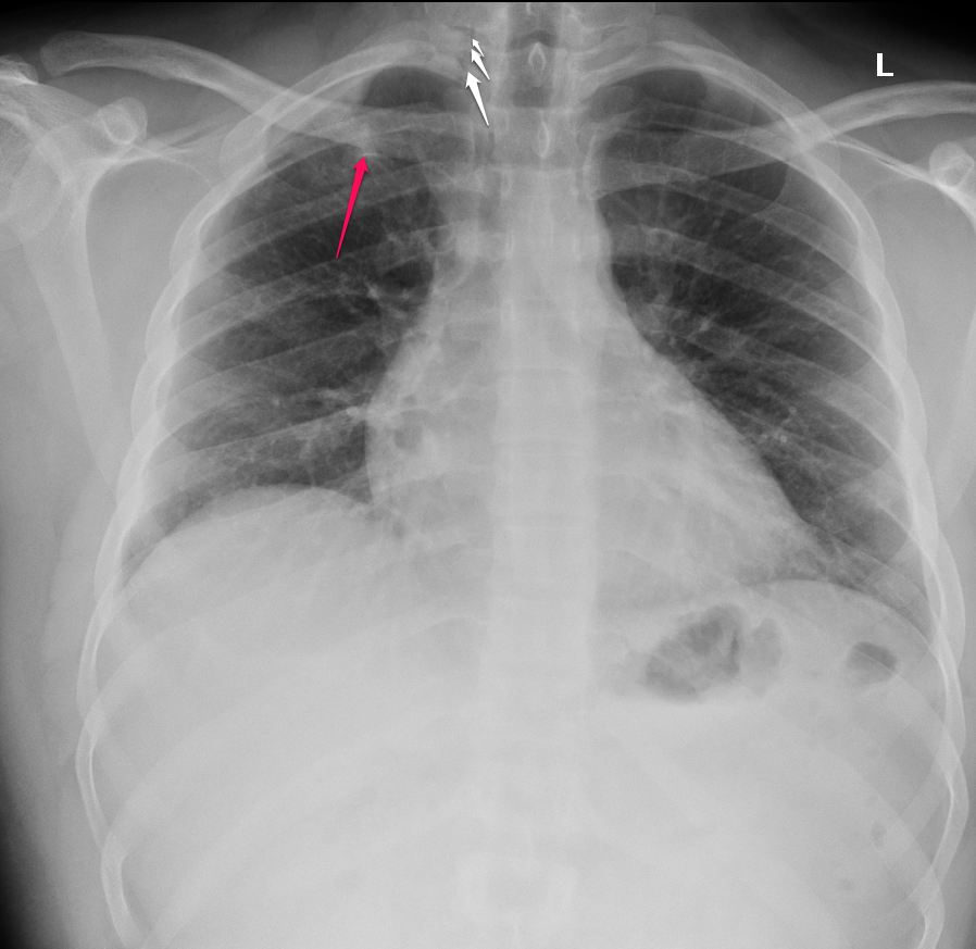
There is right 1st rib fracture with evidence of superior pneumomediastinum ( white arrows), also there is small opacity seen in the right apex ( red arrow) given the patient's history this likely represent contusion. Further evaluation by CT scan is advised.
------------------------------------------------------------------------------------------------------------


------------------------------------------------------------------------------------------------------------
Findings :


There is superior pneumomediastinum corresponding to the findings on the chest x-ray

Also there is subcutaneous emphysema.
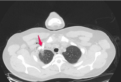
There is a displaced fracture of the first rib.

The lung apex shows an opacity in the right upper lobe adjacent to the fracture likely represent a contusion.
Subscribe to:
Posts (Atom)